Images
© Gil Hedley 2025 ~ I share these images for your study only, here and now. I entrust you to respect donors, images, and copyrights, with appreciation for these great gifts which make our studies possible. Thank you and, enjoy!
Detailed Terms of Use
Canteloupe skin: Notice the similarity of patterning between the skin of the canteloupe and the deep side of the dermis demonstrated in the next image.
Skin: This is the underside of the skin, revealing the fibrous connective tissue patterning of the dermis as it is being differentiated from the hypodermis, that fatty underlayer also known as the subcutaneous adipose, or superficial fascia.


Superficial fascia: Superficial fascia reflected and showing its fuzzy relationship to deep fascia. That white "cotton candy" is actually the dessicated (dried) "perifascial membranes" placed in tension in the dissection process revealing its "felted" collagen fiber organization.
Superficial fascia: Here is a swath of the yellow superficial fascia, also known as the hypodermis or subcutaneous adipose, at belly, differentiated and held up from deep fascia.


Superficial Fascia: At belly, differentiated and backlit. Light penetrates into our bodies and is biologically active.
Superficial Fascia portrait: ~ "Venus-Mary" ~ The entire superficial fascia (subcutaneous adipose layer) of a female form dissected as an autonomous organ and demonstrated alongside the form from which it came. I share this to transform our vision of the fatty fleece beneath our skin so we might consider it with greater appreciation, curiosity and respect.


Superficial fascia as an autonomous organ, close up: This is a great endocrine organ, a lymphoid organ, an organ of metabolism and resource, an organ of movement and sensitivity, to name a few of its many important properties essential to human health and function.
Deep Fascia: Dense, regular, fibrous deep fascia cleared of overlying superficial fascia and perifascial membranes at iliotibial band overlying vastus lateralis muscle.


Deep fascia: The deep fascia overlying the quadriceps femoris muscle is also known as the fascia lata, meaning the "broad" fascia (often misspelled "fascia latta"). Note the straight, orderly, regular array of long collageneous fibers which characterize this tissue. Note wisps of overlying perifascial membranes (aka "fuzz" or "filmy fascia") yet to be removed.
"Perifascia": Fascia lata cut and reflected showing the membranous perifascia placed in tension and looking "fuzzy." This tissue enables the differential movement between the deep fascia and the muscle tissue of the quadriceps femoris muscle. I put "perifascia" in quotes because I made up the word, which means "the fascia that's around/near the fascia" :) I also call it "filmy fascia," and used to call it "fuzz!"


Deep Fascia: Deep side of iliotibial (IT) band, demonstrating the triple-layer of dense collagenous fiber bundles, ordered at 90º relative to each other, which comprise this aspect of the fascia lata.
Perifascia, aka "fuzz": Lifting the rectus femoris, its continuous relationship with the vastus intermedius via the intervening perifascial membrane is demonstrated. The "fuzziness" is from the "felted" configuration of the collagen fiber distribution demonstrated by pulling it apart, in contrast to the regular linear patterning of the deep fascia.


Perifascia: After disrupting through dissection their fuzzy, felted continuity, we see the shiny, transparent, slippery, filmy perifascial membranes intervening between the deep side of the rectus femoris muscle (above) and the vastus intermedius (beneath). The perifascial membrane connecting them (formerly known as "fuzz") belongs there! It enables both continuity of tissues and differential movement.
Peritoneum: Respiratory diaphragm and the abdominal wall differentiated and reflected from the underlying peritoneum, the visceral fascial membrane that surrounds the abdominal viscera (organs).


Dura mater in situ: The superior sagittal sinus and the transverse sinuses are here opened to demonstrate the path of venus blood flow from the brain as it returns to the general circulation. The fact that the dura mater has two layers, both an outer periosteal layer lining the cranium and an inner meningeal layer adherent to it, create the possibility for channels for venous blood flow within them.
Right lung with 5 lobes: Although it is typical for the right lung to have three lobes, it sometimes has more, or less. In this instance there are five, due to a variation in the branching of the bronchial tree.


Brain: The human brain ex situ, with dura mater and arachnoid removed, exposing the pia mater, which is the 2-3 cell thick meningeal layer that covers the outer surface of the brain.
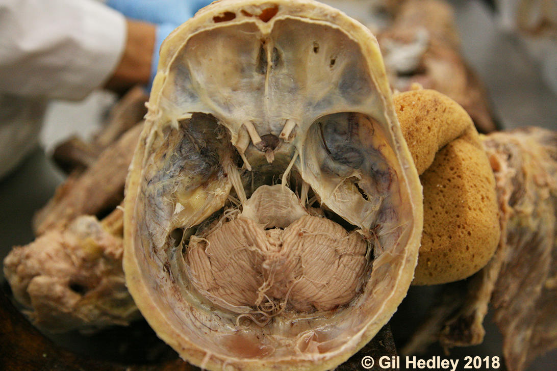
Cerebellum in situ: The brainstem has been cut and the cerebrum removed along with the meninges, including the tentorium cerebelii, to reveal the cerebellum, or "hindbrain," with numerous cut and intact cranial nerves also visible.

Cauda equina and filum terminale: The dura mater and arachnoid of the spinal cord have been incised to reveal the cauda equina, and more specifically, the filum terminale, which extends the pia mater surrounding the spinal cord beyond the conus medullaris all the way to the coccyx.
Whole dura mater intact over brain and spinal cord: Thanks to dissection course participants and dedicated somanauts Jim Donak and Jory Bell for doing the hard work to prepare this demonstration of the entire dura mater of the central nervous system ex situ, with brain and spinal cord still within.


